

")
")
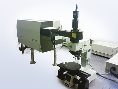
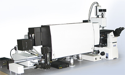
Overview:NF06 Raman Microspectroscopy system
20 years ago Tokyo Instruments has developed the industry's first apparatus for Raman imaging with spatial resolution of 200 nanometers and continues to challenge cutting-edge analysis and evaluation technology now.
Three-dimensional Raman imaging and spectroscopy can be performed with diffraction limited spatial resolution: below 200 nm in lateral (XY plane) and 500 nm in axial (Z depth) directions.
Raman spectroscopy can be combined with other analytical techniques to get extra information from the same sample area.
In order to meet the changing needs of customers, this equipment is designed to be easy expandable. The system can be combined with piezo stage, Galvano mirror scanner, stepper motor stage, cryostat, heating chamber, scanning probe microscope (SPM), streak camera, time correlation single photon counting (TCSPC) and also can be extended to nonlinear Raman spectroscopy (CARS). Customization of system configurations ensures perfect fitting for customer’s applications.
Nanofinder® software controls all system parts (opto-mechanics, scanners, detectors) as well as data acquisition, processing and analysis. Its intuitive and easy-to–handle user interface can be individually adapted to customized algorithms of system operation and data manipulation.
Demo measurements to confirm the system efficiency for customers' samples can be done. Many customers refer to data obtained with DEMO measurements of their samples as “very clear”.
After –sales maintenance support is also available.
Features
- Non-destructive sample measurement: because of extremely high system sensitivity the laser power on the sample can be reduced. Sample damage or modification is minimized.
- Extremely high spatial resolution: 200 nm (excitation laser 488 nm, oil immersion 100X microscope objective lens)
- Extremely high spectral resolution: 0.22 cm-1/CCD pixel (at wavelength 550 nm, echelle diffraction grating)
- Versatility: high flexible platform can be combined with various analytical instruments/methods
Applications
- Structural analysis and quality evaluation of carbon material:graphite, diamond, carbon black, carbon nanotube (CNT), fullerene, diamond like carbon (DLC) film, graphene etc.
- Analysis and quality evaluation of solar cell material:copper indium gallium selenide (CIGS), perovskite, quantum dots
- Detection and analysis of minute foreign substances on submicron scale (1/10,000 of a millimeter) or less
- Structural analysis and quality evaluation of lithium ion (Li-Ion) battery material
- Structural analysis and quality evaluation of organic EL (Electro Luminescence) material
- Structural analysis and quality evaluation of power device materials such as silicon carbide (SiC: compound semiconductor material composed of silicon (Si) and carbon (C))
- Analysis of a wide range of substances: gas, liquid or solid
- Analysis and evaluation of very complex biological samples, such as living organisms and human tissues
Software
Newly developed options support increased requirements from advanced researchers
Newly developed options described below are supplied with new systems, but also can be installed in previous Nanofinder®30 models. Consult with us for a possibility to upgrade your system.
Spectrometer update:
-Higher spectral resolution due to new 810 mm focal distance spectrometer
-Wider universality due to increased number of simultaneously mounted detectors
New type of 520 mm focal length spectrometer
with 3 exit ports
In addition to standard 520 mm focal length spectrometer, now it is possible to select a spectrometer with focal length 810 mm. As a result, about 1.5 times higher spectral resolution became available. Newly updated 520 mm focal length spectrometer and new 810 mm focal length spectrometer are equipped with 3 exit ports, so up to 3 different detectors can be installed simultaneously.
Fine tuning of Raman spectrometer for each sample measurement in optimal conditions: up to 5 simultaneously installed excitation lasers
In order to optimize Raman signals excitation conditions and reduce sample fluorescence level, up to five excitation lasers, operating at different wavelengths can be mounted to the system simultaneously. As a result, higher quality Raman spectra can be obtained. Laser switching (including switching of edge-filters, polarization controlling waveplates, laser shutters) can be done fully automatic by system configuration control from software.
Wider wavelength range for Raman spectroscopy: from Ultraviolet to Near Infrared
With new option for motorized optics exchange in the detection channel, the Raman detection optical path can be optimized for operation in three wavelength ranges from ultraviolet to near infrared (UV - VIS - NIR). The combination of 4 different diffraction gratings, mounted on motorized turret with 3 spectrometer exit ports for different detectors ensures optimal signal detection from UV to NIR spectral range.
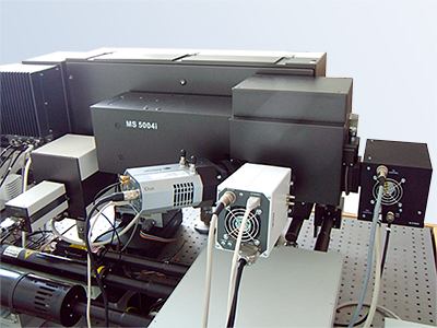
Automatic wavelength calibration (auto calibration)
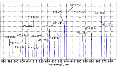
※ Rich spectrum of Ne lamp ensures high calibration accuracy
New spectra calibration option ensures always accurate wavelength calibration of the measured Raman spectra, indispensable for correct data interpretation. This motorized option introduces a neon (Ne) lamp in the optical detection path of the system. Calibration is done automatically when wavelength or diffraction grating are changed. The user can also call the calibration function any time to check accuracy of the measured data.
New mapping abilities
"Fast Mapping"
algolithm can decrease the measurement time up to 1~2ms/point.
"Large Area mapping"
applies "Fast Mapping" to stepper motor stages for sample sizes of a few cm order.
"Accumulation mapping"
automatically accumulates scanned Raman images until appropriate image quality is obtained.
"Macro spot"
sweeps the laser beam within user defined area with high speed over the sample surface to avoid damage and collect signals from full area. Can be applied to a signal "Macro spot" measurement and for mapping.
"MultiArea"
mapping algorithm provides a possibility to proceed automatical mapping in several pre-determined areas on the sample with different mapping steps and signal accumulation exposures. It also works in combination with stepper motor stage and piezo-stage, providing a possibility to get large mapping range and high spatial resolution. Configuration of selected areas can be saved and applied to the next samples.
Autofocus option for tracing of rough surfaces during 2D mapping procedure
With a confocal laser unit and the newly developed software option for reflected laser signal digital feedback, the autofocus function for surface tracing becomes possible. Even in conditions of microscope focus drift, sample inclination or rough surfaces, clear 2D-data exactly from the sample surface can be obtained. Since the confocal laser unit is independent of the Raman detection system, there is no influence on the Raman signal sensitivity or the spectrum quality.
Raman spectroscopy from <5cm-1
The new Low Frequency Raman option with motorized or manually resettable filters provides a possibility to obtain Raman spectra and even Raman imaging from 5cm-1. Both: Stokes and anti-Stokes spectra can be obtained simultaneously.
Examples of system expansion "+ α"
Expansion example 1.
Automatic piezo-stage, Galvano mirror scanner or stepper motor stage →Raman mapping in wide range at high speed · Raman imaging
Expansion example 2.
Scanning probe microscope (SPM) →Raman image and high resolution surface topography (and physical properties) from exactly the same sample area (even simultaneously),TERS
Expansion example 3.
Cryostat or heating chamber →Raman spectroscopy under environmental control
Expansion example 4.
Pulsed laser and streak camera or Time Correlated Single Photon Counter (TCSPC) →Fluorescence lifetime measurement, fluorescence intensity and lifetime imaging
Customers' special equipment (hardware and software) Unique system for customers' needs.
Examples of Nanofinder®30A Raman Microspectroscopy systems with extended functionality
Nanofinder®30A configuration for bio researchers
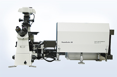
For bio applications the Nanofinder®30A system can be equipped with an inverted microscope and a Galvano mirror scanner (to keep the sample in resting state and move the laser beam for confocal imaging).
Fluorescence Lifetime Imaging Microscopy (FLIM) extension
Fluorescence lifetime measurement can be realized by combining Nanofinder®30A with ultrashort pulsed lasers such as Ti:sapphire laser or a picosecond semiconductor laser with a time correlated single photon counter (TCSPC) or a streak camera detector. Fluorescence intensity and lifetime images are obtained in 3D with high spatial resolution and high sensitivity.
Observations of intracellular molecules dynamics in biology or carrier dynamics in semiconductor materials are examples of possible applications. Temperature control with heating/cooling chamber or cryostat is also available. Sample imaging can be realized by moving the laser beam with a Galvano mirror scanner.
* Custom-made fluorescence, Photoluminescence (PL), Photoluminescence excitation (PLE) measurement systems are also acceptable
Nanofinder®30A extension for CARS Microscope
Strong Raman signals generated in the non-linear CARS (Coherent Anti-Stokes Raman Scattering) effect can be obtained in very short acquisition time and, hence, fast mapping can be realized. Ultrashort laser pulses are required for generating CARS signals from the sample.
Since the wavelength of CARS signals is shorter than the excitation laser wavelength there is no influence from sample fluorescence background. Sample preparation, such as fluorescent labeling, is not required for obtaining fast 3D CARS images
Examples of obtained mapping data
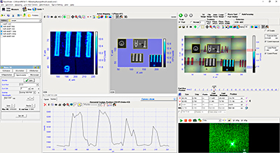
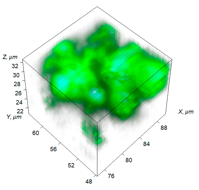
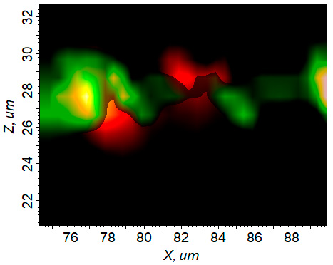
Raman spectroscopic analysis of the positive electrode of a lithium battery
The performance of Li-ion batteries deteriorates with time. The main cause of deterioration is irreversible change of the positive electrode material LiCoO2 into electrochemically inactive Co3O4. Raman imaging spectroscopy can clearly distinguish these materials and, hence, makes it possible to study the process of battery degradation with the target of the battery life prolongation.
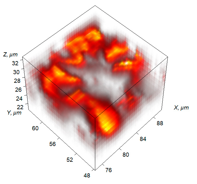
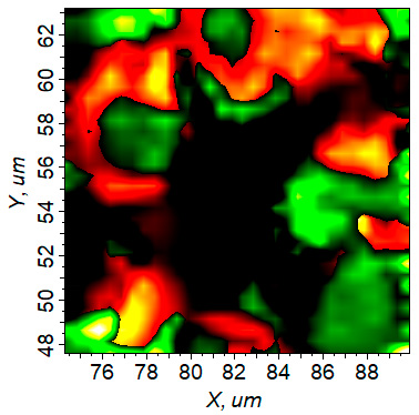
Red: LiCoO2 (normally operating parts) distribution.
Green: Co3O4(degraded parts) distribution.
Red: LiCoO2 (normally operating parts) distribution.
Green: Co3O4(degraded parts) distribution.
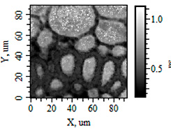
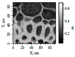
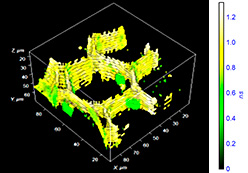
Three-dimensional fluorescence lifetime image
Fluorescence Lifetime Imaging Microscopy (FLIM) with a Time Correlated Single Photon Counter (TCSPC) provides a possibility to realize fluorescence intensity and lifetime imaging. The unique feature of the Nanofinder®30A FLIM extension is 3D fluorescence lifetime imaging with high sensitivity and high spatial resolution. Superfast FLIM data fitting (multi-exponential (up to 3) overlapped fluorescent decay curves) is ensured by parallel CPU and GPU processing.
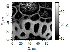
the 1st decay component
the 1st decay component
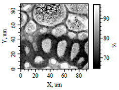
the 2nd decay component
the 2nd decay component
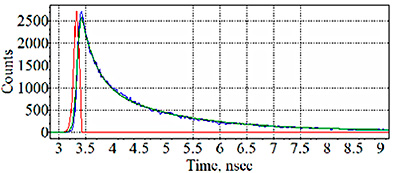
with up to 3 exponential components
Adobe Reader is required to view the downloaded PDF files.
Please download it from the left banner and install it.
| Last Updated | Update Information | Size | Download |
|---|