


Overview
Nanofinder®FLEX2 is the 2nd generation of fiber type 3D Confocal Raman microspectroscopy systems Nanofinder®FLEX series. Keeping the compactness and flexibility of fiber type layout, it brings new level of simplicity and comfort for operation with two switchable lasers. Another new feature of this device is easy and reproducible operation mode switch: High Resolution (Spectral & Spatial) or High Throughput. FLEX2 can be installed on standard up-right microscope for operation with step-motor or piezo-stage scanner or on open space granite frame microscope for combining with AFM and SNOM systems. Transmission and Inverted microscope configurations are also available.
“Line Scan option” – Safe measurement for sensitive samples
・Focusing laser beam in line to decrease power density and prevent sample damage.
・Simultaneous collection of Raman spectra from many points along the laser line.
・Easy switching between traditional Point Scan and innovative Line Scan modes with integrated slider.
※Available for:Nanofinder®FLEX2 ・Nanofinder®FLEX(soon)
Application example: Li-Ion batteries
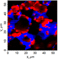
Raman intensity image of LiCoO2 (red ) and
Carbon (blue) distribution in X-Y plane
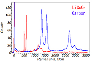
LiCoO2 and Carbon spectra , averaged (1x1µm)
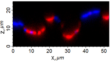
Raman intensity image of LiCoO2 (red) and Carbon (blue ) distribution in X-Z plane
Laser power on sample: 1.7 mW
Number of spectra: ~21,000
Mapping time: ~20 min
Sample courtesy of Kanamura Laboratory,
Faculty of Urban Environmental Sciences,
Tokyo Metropolitan University.
“Large Area Scan option” – Fast mapping of big samples
・Raman mapping with step motor stage of millimeter, centimeter or even larger scanning area.
・Fast scan by synchronized constant speed sample movement and data collection.
・Easy switching between point scan and fast scan.
・Combinable with “Line Scan” option.
※Stepping motor stage is required for this scan option.
Application example 1: Large test grid
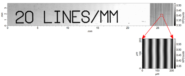
Size: 27.5 mm X 3.85 mm
Number of spectra: 218,827 (2,751x77)
Mapping time: ~25 min (~7.2 ms/point)
Application example 2: Pharmaceutical tablet
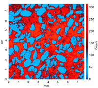
Raman intensity Image of
Aspirin (blue) and Paracetamol (red)
distribution.
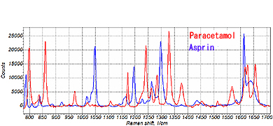
Aspirin and Paracetamol spectra,
40xNA0.6, exposure 1 min
Size: 7.75 mm X 7.75 mm
Number of spectra: 24,025 (155x155)
Mapping time: 50 min (~125 ms/point)
| Number of lasers | 2 (standard set 532 nm, 785 nm). Optional laser wavelength: 355, 473, 488, 632.8, 830, 976 nm. |
| Detection fiber bundle: 2 fiber cores | -50 m core for High Resolution (HR) mode; -105 m core for High Throughput (HT) mode. Optional fibers available. |
| Standard scanner - piezo-stage X-Y-Z | -100 m travel range; -5 nm position repeatability. Optional: other travel ranges, step-motor scanner, AFM scanner. |
| Laser | 532 nm | 785 nm |
| Wavenumber range(cm-1) | 50~4000 | 40~3000 |
| Dispersiona)(cm-1/pix) | 1.3(at 546 nm) | 0.49(at 812 nm) |
| Spectral Resolutiona),b)FWHM,typ(max)(cm-1) | <2(2.6)(at 546 nm) | <0.75(1)(at 812 nm) |
| Spatial resolutionb),c)X-Y(nm) | <350 | <500 |
| Spatial resolutionb),c)Z(nm) | <900 | <900 |
a) with spectrometer f=350 mm, grating 1800G/mm, entrance slit 30 m, CCD pixel 26 m.
b) fiber 50 m core ("High Resolution" confocal mode)
c) with objective lens 100X NA=0.95
| Size | Microscope with "Flex2" Raman unit footprint ● Olympus microscope frame:270×380 mm ● Free Space Granite frame:390×580 mm |
| Standard 350 mm spectrometer footprint: 510×395 mm | |
| Power consumption: 100 V~15 A or 220 V~7.5 A |
Advanced Software
■ Full system control and 1-2-3D data analysis and visualization;
■ Full Raman spectrum saving in every mapping point;
■ Simultaneous multidetectors readout;
■ Possibility to select mapping area on TV-CCD microscopic sample image;
■ Fast scanning algorithm;
■ Spectra stitching;
■ Throughput correction;
■ Macro-spot mapping;
■ Image accumulation;
■ Spectrum fitting with up to 5 Lorentzian or Gaussian curves;
■ Deconvolution processing for 1D or 2D data
■ Simultaneous AFM topography and Raman spectroscopy,same area and TERS functions.
Adobe Reader is required to view the downloaded PDF files.
Please download it from the left banner and install it.
| Last Updated | Update Information | Size | Download |
|---|